Higher Total Omega-3 Intake is Associated with Less Vascular Calcification in Older Women
This article at a glance 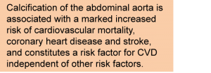 Interestingly, research in the last few years has generated a shift in the understanding of the vascular calcification process from a passive mineralization with deposition of calcium phosphate crystals within tissues to a dysregulated but active formation of true bone-like structures. This new insight views calcification within the arterial vasculature as a result of the improper activation of cells and hormones that normally regulate skeletal bone formation. This comprises the differentiation of vascular smooth muscle cells to acquire a mineralizing chondrocyte-like function. These osteoblast-like cells produce calcium-containing vesicles and a matrix of bone collagen and noncollagenous proteins that can then mineralize if the balance of pro-mineralizing factors outweighs inhibitory factors. Also, monocyte-derived cells with osteoclast-like activity are present in atherosclerotic calcified structures. In contrast to medial calcification, intimal biomineralization and deregulated bone biology has been shown to be closely associated with chronic inflammation, which is also important to the atherosclerotic process. Previous interpretations that calcification of atherosclerotic plaques confers a stabilizing effect on atherosclerotic plaques that become less susceptible to rupturing and subsequent thrombus formation have in recent years become increasingly challenged by new views. This evolving topic of research demonstrates that calcification may also be involved in promoting further plaque remodelling, and is intimately involved in destabilization of the fibrous cap that covers advanced atherosclerotic plaques through the seeding of microcalcified structures from matrix vesicles. Mineralization of the arterial vasculature is also considered an intermediate step in the progression to either weakening of the arterial wall (potentially leading to aneurism) or to vascular stenosis with more extensive calcification and arterial obstruction. Calcification of arterial vessels other than coronary or cerebral vessels was suggested to have predictive relevance for CVD death 30 years ago. Calcification of the abdominal aorta is associated with a marked increased risk of cardiovascular mortality, coronary heart disease and stroke, and constitutes a risk factor for CVD independent of other risk factors. Aortic calcification is positively associated with age, with older people having more severe calcification of the abdominal aorta. Other factors associated to calcification of the aorta are hypertension, smoking, the presence of circulating markers of inflammation (CRP) and bone metabolism (osteoprotegerin, osteopontin), and disturbed lipoprotein and lipid metabolism. Aortic calcification is a common feature in patients with diabetes, chronic kidney disease/renal failure, and osteoporosis.
 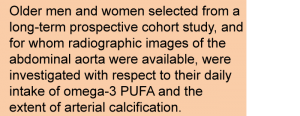 Given the involvement of inflammation in the misdirected biomineralization process of the intima, and the etiological role of vascular inflammation in the atherosclerotic process, it may be possible to modify or redirect the calcification process and associated arterial complications. Results from animal studies have suggested that vascular calcification can be reduced by increased omega-3 LCPUFA intake. The potential relationship between omega-3 status or omega-3 PUFA dietary intake and aortic calcification in humans has never been assessed. A recent report on the results of a large and long-lasting prospective cohort study has now investigated if dietary alpha-linolenic acid (ALA), EPA plus DHA, or total omega-3 PUFA were associated with the development of abdominal aortic calcification. The study focused on arterial calcification in older people, where the calcification load is highest. The study results were reported by Shang and colleagues from the Faculty of Medicine, University of Melbourne, Australia, and several other institutes in Melbourne and Delhi, India. The study examined 312 selected study participants from the >41000 adults that enrolled at baseline between 1990 and 1994, at age 45-64 years old, for whom readable lateral thoraco-lumbar radiographs were available at follow-up and whose dietary intake characteristics had been determined at study onset. Dietary intake was again assessed at follow-up when the selected study subjects were on average 18 ± 1 years older. Dietary intake was measured using food-frequency questionnaires designed specifically for the Melbourne Collaborative Cohort Study, the prospective cohort study from which the 312 selected participants were selected. Of note, only subjects with a calcium intake greater than 1300 mg/d or below 500 mg/d were recruited. Information on dietary habits including use of milk, sugar, supplements, and type of oils and other fats was collected. The intake of omega-3 PUFA (total, ALA, EPA, DHA) was calculated using an Australian food and compositional database.
  The degree of vascular calcification of the descending aorta was measured at follow-up at the level of the thoraco-lumbar region of the spine. Two complementary non-invasive imaging techniques for measuring mineralized tissue (bone) density were used: thoraco-lumbar lateral X-ray radiography and dual-energy X-ray absorptiometric imaging (DXA). A semi-quantitative composite and summed score of calcified deposits in the abdominal aortic vascular wall was made by independent and trained evaluators to grade the severity of arterial calcification at the level of the first through fourth lumbar vertebrae. Males and females were found to have significant differences in energy-adjusted daily intake of omega-3 PUFA, and baseline characteristics were therefore evaluated by gender according to tertiles of total omega-3 PUFA intake. Men in the highest tertile intake of total omega-3 PUFA had significantly higher energy intake, as well as higher intake of fiber ALA, EPA plus DHA, fruit vegetables, fish, meat and nuts. Women in the highest tertile of total omega-3 intake had significantly higher energy intake, as well as higher ALA, EPA plus DHA, and vegetable intake. Women in the lowest tertile had significantly lower fish and meat intake. Calcium intake was significantly lower in the second tertile in both men and women, with no difference between higher calcium intake in lowest and highest tertiles of total omega-3 PUFA intake. No other dietary or anthropometric differences were measurable. No significant difference in the extent of calcification of the abdominal aorta was found between men with low, intermediate or high total omega-3 PUFA daily intake. However, a significantly higher proportion of women with the highest omega-3 PUFA daily intake had no calcification. Accordingly, fewer women had high or moderate calcification scores when measured by radiography. When measured by DXA, a significantly lower proportion of women in the lowest tertile of total omega-3 intake were free of aortic calcification. In older women there was a statistically significant inverse relation between energy-adjusted ALA intake and calcification after adjustment for a number of potential confounders (age, smoking, physical activity, BMI, systolic and diastolic blood pressure, plasma cholesterol, total energy and calcium intakes). An inverse association was also observed for total omega-3 PUFA intake and aortic calcification in women that was also apparent in regression analyses adjusted for age only. The strength of the associations varied somewhat depending on the imaging technique used, and was stronger after adjustment for a larger number of potential confounders. For EPA plus DHA intake no significant association was found (0.42 g/d median intake in tertile 3). There were no associations between total omega-3 PUFA, ALA intake, or EPA plus DHA (median intake in highest tertile, 0.35 g/d) in men. Changes in tertile of energy-adjusted ALA, EPA/DHA, or total omega-3 PUFA intake over the eighteen-year study period were not associated with the level of abdominal aortic calcification. The results of this study indicate that baseline intake of ALA and total omega-3 PUFA in older women is associated with lower aortic calcification severity over an eighteen-year period.
 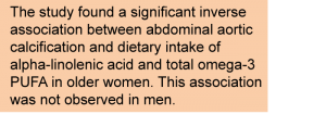 Men have a higher prevalence of arterial calcification than women, but calcification of the abdominal aorta is the most common arterial site for calcification in women. A potentially detrimental relation between abdominal aortic calcification and bone loss has been reported in older women, but appears absent in men. This study evaluated men and women with median intakes of EPA plus DHA that were relatively low, and accounted for a small proportion of total omega-3 LCPUFA. It is possible that at higher daily EPA/DHA intake, an association with lower calcification severity may also become apparent. Men in this study were not meeting the recommended daily intake level for ALA (in Australia). No relationship between low or high calcium intake and calcification of the abdominal aorta was found in this study. The results of this study indicate for the first time that biomineralization of the abdominal aorta as an independent risk factor for cardiovascular disease is inversely associated with dietary intake of omega-3 PUFA and ALA, albeit only in older women. Whether in older men arterial calcification may be modifiable or is related to dietary omega-3 PUFA intake at higher intake levels will require further research. As a potentially modifiable outcome by diet, addressing calcification of the abdominal aorta may furthermore have important implications for bone health in older women. Shang X, Sanders KM, Scott D, Khan B, Hodge A, Khan N, English DR, Giles GG, Ebeling PR. Dietary alpha-linolenic acid and total omega-3 fatty acids are inversely associated with abdominal aortic calcification in older women, but not in older men. J. Nutr. 2015;145(8):1778-1786. [PubMed] Worth Noting Allison MA, Criqui MH, Wright CM. Patterns and risk factors for systemic calcified atherosclerosis. Arterioscler Thromb. Vasc. Biol. 2004;24(2):331-336. [PubMed] Bobryshev YV, Killingsworth MC, Lord RS, Grabs AJ. Matrix vesicles in the fibrous cap of atherosclerotic plaque: possible contribution to plaque rupture. J. Cell. Mol. Med. 2008;12(5B):2073-2082. [PubMed] Doherty TM, Asotra K, Fitzpatrick LA, Qiao JH, Wilkin DJ, Detrano RC, Dunstan CR, Shah PK, Rajavashisth TB. Calcification in atherosclerosis: bone biology and chronic inflammation at the arterial crossroads. Proc. Natl. Acad. Sci. USA 2003;100(20):11201-11206. [PubMed] Dual-energy X-ray absorptiometry. Radiology Info for patients. http://www.radiologyinfo.org/en/info.cfm?pg=dexa Ewence AE, Bootman M, Roderick HL, Skepper JN, McCarthy G, Epple M, Neumann M, Shanahan CM, Proudfoot D. Calcium phosphate crystals induce cell death in human vascular smooth muscle cells: a potential mechanism in atherosclerotic plaque destabilization. Circ. Res. 2008;103(5):e28-34. [PubMed] Giles GG, English DR. The Melbourne Collaborative Cohort Study. IARC Sci. Publ. 2002;156:69-70. [PubMed] Hutcheson JD, Maldonado N, Aikawa E. Small entities with large impact: microcalcifications and atherosclerotic plaque vulnerability. Curr. Opin. Lipidol. 2014;25(5):327-332. [PubMed] Jayalath RW, Mangan SH, Golledge J. Aortic calcification. Eur. J. Vasc. Endovasc. Surg. 2005;30(5):476-488. [PubMed] Kiel DP, Kauppila LI, Cupples LA, Hannan MT, O'Donnell CJ, Wilson PW. Bone loss and the progression of abdominal aortic calcification over a 25 year period: the Framingham Heart Study. Calcif. Tissue Int. 2001;68(5):271-276. [PubMed] Nestel P, Clifton P, Colquhoun D, Noakes M, Mori TA, Sullivan D, Thomas B. Indications for omega-3 long chain polyunsaturated fatty acid in the prevention and treatment of cardiovascular disease. Heart Lung Circ. 2015;24(8):769-779. [PubMed] Reaven PD, Sacks J. Reduced coronary artery and abdominal aortic calcification in Hispanics with type 2 diabetes. Diabetes Care 2004;27(5):1115-1120. [PubMed] Virmani R, Joner M, Sakakura K. Calcification. Arterioscler. Thromb. Vasc. Biol. 2015;34:1329-1332. [PubMed] Walsh CR, Cupples LA, Levy D, Kiel DP, Hannan M, Wilson PW, O'Donnell CJ. Abdominal aortic calcific deposits are associated with increased risk for congestive heart failure: the Framingham Heart Study. Am. Heart J. 2002;144(4):733-739. [PubMed] Wilson PW, Kauppila LI, O'Donnell CJ, Kiel DP, Hannan M, Polak JM, Cupples LA. Abdominal aortic calcific deposits are an important predictor of vascular morbidity and mortality. Circulation 2001;103(11):1529-1534. [PubMed] Witteman JC, Kok FJ, van Saase JL, Valkenburg HA. Aortic calcification as a predictor of cardiovascular mortality. Lancet 1986;2(8516):1120-1122. [PubMed] Yamanouchi D, Takei Y, Komori K. Balanced mineralization in the arterial system: possible role of osteoclastogenesis/osteoblastogenesis in abdominal aortic aneurysm and stenotic disease. Circ. J. 2012;76(12):2732-2737. [PubMed]
- Calcification of the abdominal aorta is a predictor of cardiovascular disease (CVD), independent of other CVD risk factors.
- This study addressed whether vascular calcification development in adults followed over an 18-year period displays a relation to dietary intake of omega-3 polyunsaturated fatty acids.
- The study reports that higher alpha-linolenic acid and total omega-3 fatty acid intake is associated with reduced severity of aortic calcification in women. This association was not found in men.
- The results may help identify additional patient groups that are at risk for CVD.
Interestingly, research in the last few years has generated a shift in the understanding of the vascular calcification process from a passive mineralization with deposition of calcium phosphate crystals within tissues to a dysregulated but active formation of true bone-like structures. This new insight views calcification within the arterial vasculature as a result of the improper activation of cells and hormones that normally regulate skeletal bone formation. This comprises the differentiation of vascular smooth muscle cells to acquire a mineralizing chondrocyte-like function. These osteoblast-like cells produce calcium-containing vesicles and a matrix of bone collagen and noncollagenous proteins that can then mineralize if the balance of pro-mineralizing factors outweighs inhibitory factors. Also, monocyte-derived cells with osteoclast-like activity are present in atherosclerotic calcified structures. In contrast to medial calcification, intimal biomineralization and deregulated bone biology has been shown to be closely associated with chronic inflammation, which is also important to the atherosclerotic process. Previous interpretations that calcification of atherosclerotic plaques confers a stabilizing effect on atherosclerotic plaques that become less susceptible to rupturing and subsequent thrombus formation have in recent years become increasingly challenged by new views. This evolving topic of research demonstrates that calcification may also be involved in promoting further plaque remodelling, and is intimately involved in destabilization of the fibrous cap that covers advanced atherosclerotic plaques through the seeding of microcalcified structures from matrix vesicles. Mineralization of the arterial vasculature is also considered an intermediate step in the progression to either weakening of the arterial wall (potentially leading to aneurism) or to vascular stenosis with more extensive calcification and arterial obstruction. Calcification of arterial vessels other than coronary or cerebral vessels was suggested to have predictive relevance for CVD death 30 years ago. Calcification of the abdominal aorta is associated with a marked increased risk of cardiovascular mortality, coronary heart disease and stroke, and constitutes a risk factor for CVD independent of other risk factors. Aortic calcification is positively associated with age, with older people having more severe calcification of the abdominal aorta. Other factors associated to calcification of the aorta are hypertension, smoking, the presence of circulating markers of inflammation (CRP) and bone metabolism (osteoprotegerin, osteopontin), and disturbed lipoprotein and lipid metabolism. Aortic calcification is a common feature in patients with diabetes, chronic kidney disease/renal failure, and osteoporosis. Given the involvement of inflammation in the misdirected biomineralization process of the intima, and the etiological role of vascular inflammation in the atherosclerotic process, it may be possible to modify or redirect the calcification process and associated arterial complications. Results from animal studies have suggested that vascular calcification can be reduced by increased omega-3 LCPUFA intake. The potential relationship between omega-3 status or omega-3 PUFA dietary intake and aortic calcification in humans has never been assessed. A recent report on the results of a large and long-lasting prospective cohort study has now investigated if dietary alpha-linolenic acid (ALA), EPA plus DHA, or total omega-3 PUFA were associated with the development of abdominal aortic calcification. The study focused on arterial calcification in older people, where the calcification load is highest. The study results were reported by Shang and colleagues from the Faculty of Medicine, University of Melbourne, Australia, and several other institutes in Melbourne and Delhi, India. The study examined 312 selected study participants from the >41000 adults that enrolled at baseline between 1990 and 1994, at age 45-64 years old, for whom readable lateral thoraco-lumbar radiographs were available at follow-up and whose dietary intake characteristics had been determined at study onset. Dietary intake was again assessed at follow-up when the selected study subjects were on average 18 ± 1 years older. Dietary intake was measured using food-frequency questionnaires designed specifically for the Melbourne Collaborative Cohort Study, the prospective cohort study from which the 312 selected participants were selected. Of note, only subjects with a calcium intake greater than 1300 mg/d or below 500 mg/d were recruited. Information on dietary habits including use of milk, sugar, supplements, and type of oils and other fats was collected. The intake of omega-3 PUFA (total, ALA, EPA, DHA) was calculated using an Australian food and compositional database. The degree of vascular calcification of the descending aorta was measured at follow-up at the level of the thoraco-lumbar region of the spine. Two complementary non-invasive imaging techniques for measuring mineralized tissue (bone) density were used: thoraco-lumbar lateral X-ray radiography and dual-energy X-ray absorptiometric imaging (DXA). A semi-quantitative composite and summed score of calcified deposits in the abdominal aortic vascular wall was made by independent and trained evaluators to grade the severity of arterial calcification at the level of the first through fourth lumbar vertebrae. Males and females were found to have significant differences in energy-adjusted daily intake of omega-3 PUFA, and baseline characteristics were therefore evaluated by gender according to tertiles of total omega-3 PUFA intake. Men in the highest tertile intake of total omega-3 PUFA had significantly higher energy intake, as well as higher intake of fiber ALA, EPA plus DHA, fruit vegetables, fish, meat and nuts. Women in the highest tertile of total omega-3 intake had significantly higher energy intake, as well as higher ALA, EPA plus DHA, and vegetable intake. Women in the lowest tertile had significantly lower fish and meat intake. Calcium intake was significantly lower in the second tertile in both men and women, with no difference between higher calcium intake in lowest and highest tertiles of total omega-3 PUFA intake. No other dietary or anthropometric differences were measurable. No significant difference in the extent of calcification of the abdominal aorta was found between men with low, intermediate or high total omega-3 PUFA daily intake. However, a significantly higher proportion of women with the highest omega-3 PUFA daily intake had no calcification. Accordingly, fewer women had high or moderate calcification scores when measured by radiography. When measured by DXA, a significantly lower proportion of women in the lowest tertile of total omega-3 intake were free of aortic calcification. In older women there was a statistically significant inverse relation between energy-adjusted ALA intake and calcification after adjustment for a number of potential confounders (age, smoking, physical activity, BMI, systolic and diastolic blood pressure, plasma cholesterol, total energy and calcium intakes). An inverse association was also observed for total omega-3 PUFA intake and aortic calcification in women that was also apparent in regression analyses adjusted for age only. The strength of the associations varied somewhat depending on the imaging technique used, and was stronger after adjustment for a larger number of potential confounders. For EPA plus DHA intake no significant association was found (0.42 g/d median intake in tertile 3). There were no associations between total omega-3 PUFA, ALA intake, or EPA plus DHA (median intake in highest tertile, 0.35 g/d) in men. Changes in tertile of energy-adjusted ALA, EPA/DHA, or total omega-3 PUFA intake over the eighteen-year study period were not associated with the level of abdominal aortic calcification. The results of this study indicate that baseline intake of ALA and total omega-3 PUFA in older women is associated with lower aortic calcification severity over an eighteen-year period. Men have a higher prevalence of arterial calcification than women, but calcification of the abdominal aorta is the most common arterial site for calcification in women. A potentially detrimental relation between abdominal aortic calcification and bone loss has been reported in older women, but appears absent in men. This study evaluated men and women with median intakes of EPA plus DHA that were relatively low, and accounted for a small proportion of total omega-3 LCPUFA. It is possible that at higher daily EPA/DHA intake, an association with lower calcification severity may also become apparent. Men in this study were not meeting the recommended daily intake level for ALA (in Australia). No relationship between low or high calcium intake and calcification of the abdominal aorta was found in this study. The results of this study indicate for the first time that biomineralization of the abdominal aorta as an independent risk factor for cardiovascular disease is inversely associated with dietary intake of omega-3 PUFA and ALA, albeit only in older women. Whether in older men arterial calcification may be modifiable or is related to dietary omega-3 PUFA intake at higher intake levels will require further research. As a potentially modifiable outcome by diet, addressing calcification of the abdominal aorta may furthermore have important implications for bone health in older women. Shang X, Sanders KM, Scott D, Khan B, Hodge A, Khan N, English DR, Giles GG, Ebeling PR. Dietary alpha-linolenic acid and total omega-3 fatty acids are inversely associated with abdominal aortic calcification in older women, but not in older men. J. Nutr. 2015;145(8):1778-1786. [PubMed] Worth Noting Allison MA, Criqui MH, Wright CM. Patterns and risk factors for systemic calcified atherosclerosis. Arterioscler Thromb. Vasc. Biol. 2004;24(2):331-336. [PubMed] Bobryshev YV, Killingsworth MC, Lord RS, Grabs AJ. Matrix vesicles in the fibrous cap of atherosclerotic plaque: possible contribution to plaque rupture. J. Cell. Mol. Med. 2008;12(5B):2073-2082. [PubMed] Doherty TM, Asotra K, Fitzpatrick LA, Qiao JH, Wilkin DJ, Detrano RC, Dunstan CR, Shah PK, Rajavashisth TB. Calcification in atherosclerosis: bone biology and chronic inflammation at the arterial crossroads. Proc. Natl. Acad. Sci. USA 2003;100(20):11201-11206. [PubMed] Dual-energy X-ray absorptiometry. Radiology Info for patients. http://www.radiologyinfo.org/en/info.cfm?pg=dexa Ewence AE, Bootman M, Roderick HL, Skepper JN, McCarthy G, Epple M, Neumann M, Shanahan CM, Proudfoot D. Calcium phosphate crystals induce cell death in human vascular smooth muscle cells: a potential mechanism in atherosclerotic plaque destabilization. Circ. Res. 2008;103(5):e28-34. [PubMed] Giles GG, English DR. The Melbourne Collaborative Cohort Study. IARC Sci. Publ. 2002;156:69-70. [PubMed] Hutcheson JD, Maldonado N, Aikawa E. Small entities with large impact: microcalcifications and atherosclerotic plaque vulnerability. Curr. Opin. Lipidol. 2014;25(5):327-332. [PubMed] Jayalath RW, Mangan SH, Golledge J. Aortic calcification. Eur. J. Vasc. Endovasc. Surg. 2005;30(5):476-488. [PubMed] Kiel DP, Kauppila LI, Cupples LA, Hannan MT, O'Donnell CJ, Wilson PW. Bone loss and the progression of abdominal aortic calcification over a 25 year period: the Framingham Heart Study. Calcif. Tissue Int. 2001;68(5):271-276. [PubMed] Nestel P, Clifton P, Colquhoun D, Noakes M, Mori TA, Sullivan D, Thomas B. Indications for omega-3 long chain polyunsaturated fatty acid in the prevention and treatment of cardiovascular disease. Heart Lung Circ. 2015;24(8):769-779. [PubMed] Reaven PD, Sacks J. Reduced coronary artery and abdominal aortic calcification in Hispanics with type 2 diabetes. Diabetes Care 2004;27(5):1115-1120. [PubMed] Virmani R, Joner M, Sakakura K. Calcification. Arterioscler. Thromb. Vasc. Biol. 2015;34:1329-1332. [PubMed] Walsh CR, Cupples LA, Levy D, Kiel DP, Hannan M, Wilson PW, O'Donnell CJ. Abdominal aortic calcific deposits are associated with increased risk for congestive heart failure: the Framingham Heart Study. Am. Heart J. 2002;144(4):733-739. [PubMed] Wilson PW, Kauppila LI, O'Donnell CJ, Kiel DP, Hannan M, Polak JM, Cupples LA. Abdominal aortic calcific deposits are an important predictor of vascular morbidity and mortality. Circulation 2001;103(11):1529-1534. [PubMed] Witteman JC, Kok FJ, van Saase JL, Valkenburg HA. Aortic calcification as a predictor of cardiovascular mortality. Lancet 1986;2(8516):1120-1122. [PubMed] Yamanouchi D, Takei Y, Komori K. Balanced mineralization in the arterial system: possible role of osteoclastogenesis/osteoblastogenesis in abdominal aortic aneurysm and stenotic disease. Circ. J. 2012;76(12):2732-2737. [PubMed]